- Received May 25, 2022
- Accepted July 06, 2022
- Publication September 23, 2022
- Visibility 5 Views
- Downloads 0 Downloads
- DOI 10.18231/j.achr.2022.043
-
CrossMark

- Citation
Signet ring cells in cervical Pap smear – Artifactual Vs Reactive-reparative Vs Malignant: A case report and review of literature
- Author Details:
-
 Simranjeet Kaur *
-
Sandeep Kumar
Introduction
SRCs have cytoplasmic large round-oval vacuole/aggregates of vacuoles pushing and compressing the nucleus to periphery. Vacuoles may contain mucin/other material/may be empty.[1] Few cases have been reported previously, as seen in association with non-neoplastic/neoplastic cervical lesions, metastatic deposits or as artifactual. So due to diagnostic dilemma and rarity of such cases being reported, we present the case along with literature review.
Practitioner points
Signet Ring Cells in cervix can be part of IUD usage or as a part of malignancy.
Artifactual SRCs in cervical Pap may many times masquerade as part of malignancy.
Supportive clinico-radiological findings and close follow-up must always be considered when SRCs are seen in the cervical sample.
Cass Report
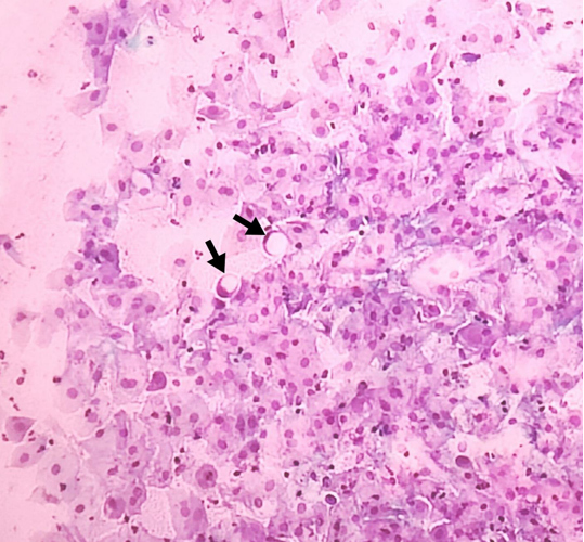
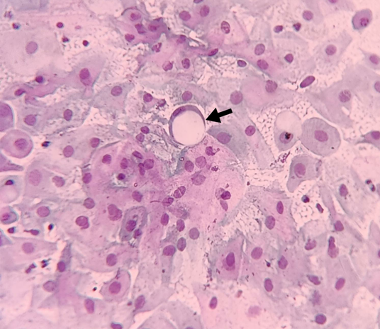
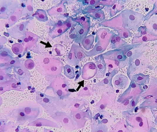
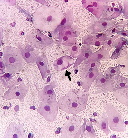
A 50 years postmenopausal female presented with itching and foul smelling discharge per vaginum and no history of IUD. Local examination showed white discharge along with inflammation. Ultrasound showed 15×16mm uterine fibroid. Cytology revealed mainly benign superficial and intermediate squamous epithelial cells and interspersed SRCs, along with neutrophils. ([Figure 1], [Figure 2]). There were other cells with single or multiple varying size vacuoles displacing nucleus. ([Figure 3], [Figure 4]). Additional workups, colposcopy showed no abnormalities. In last 1year follow-up, she has developed no new signs/symptoms. Since clinico-radiological correlation ruled out malignancy and with no IUD history, these SRCs on cervical cytology were labelled artifactual.
|
True SRCs |
False SRCs/SRLCs: Artifactual |
False SRCs: Reactive-reparative cellular changes |
|
Intracytoplasmic vacuoles containing mucin or other material |
Vacuoles are usually empty |
Vacuoles are usually empty |
|
Usually associated with underlying malignancy |
Can result from artifactual shrinkage related to fixation or procedural trauma |
Usually associated with long-term placed IUD (SRCs here are exfoliated endometrial/ endocervical columnar cells due to chronic irritation.) |
|
Nuclei correspond in morphology to that of malignant counterpart |
Nuclei appear relatively normal |
Nuclei appear relatively normal |
|
Immunohistochemistry (IHC) may be positive depending on underlying malignancy, like: positive for p16 and carcinoembryonic antigen, CK7, MUC1,5,6 |
Negative for IHC |
Negative for IHC |
|
Occasionally intracytoplasmic lumen is lined by microvilli |
Not seen |
Not seen |
|
Correlating clinico-radiological features of malignancy are present |
Correlating clinico-radiological features of malignancy are not present |
Correlating clinico-radiological features of malignancy may/may not be present |
|
Author |
Number of cases |
Malignant SRCs |
Non-malignant SRCs with H/O IUD |
Non-malignant SRCs without H/O IUD |
Diagnosis on cytology and/or histopathology |
|
Rajyaguru K.M. et al |
One |
- |
- |
Y |
HSIL (C) |
|
Daniel F.I. Kurtycz et al |
One |
- |
Y |
- |
HSIL (C) |
|
Satoshi Kawai et al |
One |
Y |
- |
- |
SRC predominant cervical adenocarcinoma (C) |
|
Kohei Hamada et al |
One |
Y |
- |
- |
SRC predominant cervical adenocarcinoma (H) |
|
Bernadette Cracchiolo et al |
One |
Y |
- |
- |
PSRCC (H) |
|
Veysel S al et al |
One |
Y |
- |
- |
PSRCC (H) |
Discussion
SRCs in cervix can be seen in non-neoplastic lesions i.e. reactive-reparative, rarely in neoplasms and artifactual as seen in present case. ([Table 1])[2], [3], [4] Daniel F.I. Kurtycz et al documented SRCs due to chronic irritation by IUD device. With absence of other morphological features to suggest High Grade Squamous Intraepithelial Lesion (HSIL), misdiagnosis was thus avoided. Hence IUD history is important.[5] There was no IUD usage in present case.
Malignancy associated SRCs, were seen on histopathology in SRC predominant cervical adenocarcinoma in a 40years female on histopathology by Kohei Hamada et al and SRC type adenocarcinoma with HPV positive full genome in a 40years female on cervical cytology by Satoshi Kawai et al.[6], [7] There were supportive clinico-radiological findings like cervical mass in 48years with large pelvic mass in lower uterine segment/cervix and in 64years females with primary signet ring cell (PSRCC) carcinoma on pan hysterectomy and endometrial/endocervical curettage, diagnosed by Bernadette Cracchiolo et al and Veysel Sal et al respectively.[8], [9] Correlation with clinico-radiological features is important to support the carcinoma diagnosis even after the presence of SRCs on microscopy. In the present case, no such pointers suggested suspicion towards malignancy. Various studies where morphological SRCs were found in cervix are tabulated in [Table 2] along with histological diagnosis.
Cases of artifactual SRCs were reported from other sites like on duodenal (strictured area) biopsy in 89years male reported by Mark Li-chang Wu. This was at first misinterpreted as SRC carcinoma. Other indicators of malignancy were ruled out. SRCs found were detached, degenerated epithelial cells from normal glands post-procedural mechanical trauma. As proposed by author, formation of soapy water bubbles from a child's toy can be analogous for artifactual SRCs production. The cell is pushed out of gland like soapy-water film bulging out from ring, when gland being assumed analogous to plastic ring, epithelial cells to soapy-water film bound by ring and procedural trauma to airflow directed through ring. After detachment from gland (ring), cells achieve spherical shape (bubbles) to achieve physical equilibrium. They arbitrarily categorised SRCs into true and false SRCs/ SRLCs (Signet ring like cells).[2]
Careful microscopic examination as well as clinico-radiological correlation must be done to rule out artifactual causes of SRCs, before labelling patient as case of PSRCC carcinoma.[[Table 2]] [5], [6], [7], [8], [9], [10]
Conclusion
Third category of SRCs as artifactual can be considered to streamline pathological causes of SRCs. Proper sampling, tissue fixation and history taking are key pointers to reach at correct diagnosis.
Conflicts of Interest
None.
Source of Funding
None.
References
- S E J C Iezzoni, Mills. Nonneoplastic endometrial signet-ring cells. Vacuolated decidual cells and stromal histiocytes mimicking adenocarcinoma. Am J Clin Pathol 2001. [Google Scholar]
- M Li-Cheng Wu, S Natarajan, KJ Lewin. Peculiar artifacts mimicking carcinoma. Arch Pathol Lab Med 2001. [Google Scholar]
- MM Wang, NR Wang, LR Deng, J Deng, SL Zhou, JX Xu. Signet-ring cell in papilloma virus type 6 and 16-related non-neoplastic squamous cell lesion. Int J Clin Exp Pathol 2016. [Google Scholar]
- YH Kim, SJ Lee, SU Lee, HI Sun, Y Kwang Il, KJ Hwi. Primary signet ring cell carcinoma of the uterine cervix. Medicine 2021. [Google Scholar] [Crossref]
- FI Daniel, PN Kurtycz, NA Staats, M Young, TJ Bibbo, MU Prey, R Nayar, DC Wilbur. Non-neoplastic Findings. The Bethesda System for Reporting Cervical Cytology. Definitions, Criteria and Explanatory notes. 3rd edn. 2015. [Google Scholar]
- K Hamada, T Baba, A Takaori, R Murakami, A Horie, K Abiko. Primary signet ring cell carcinoma of uterine cervix and related disease: two case reports and a review. Int Cancer Conf J 2019. [Google Scholar]
- S Kawai, Y Torii, I Kukimoto, T Fujii. A case of primary signet-ring cell carcinoma of the cervix containing full genome of human papillomavirus 16. Indian J Pathol Microbiol 2019. [Google Scholar]
- B Cracchiolo, T Kuhn, D Heller. Primary signet ring cell adenocarcinoma of the uterine cervix - A rare neoplasm that raises the question of metastasis to the cervix. Gynecol Oncol Rep 2016. [Google Scholar] [Crossref]
- V Sal, I Kahramanoglu, H Turan, N Tokgozoglu, T Bese, O Aydin. Primary signet ring cell carcinoma of the cervix: A case report and review of the literature. Int J Surg Case Rep 2016. [Google Scholar] [Crossref]
- KM Rajyaguru, A Zakuan, SI Shahruddin. A Rare Presentation of Signet Ring Squamous Cells in Ectocervical Squamous Epithelium Causing Diagnostic Difficulty in Reporting Cervical Intra-Epithelial Neoplasia. BJMMR 2016. [Google Scholar]